

Q Fever menu
Veterinarians
This area has more details on infection with Coxiella burnetii / Q Fever which is an important zoonotic disease with a wide geographical distribution.
Positive Coxiella burnetti PCR results should be reported in Ireland to the Department of Agriculture. More information can be found here.
More information about reporting to the department will be shared here shortly.
Coxiella burnetii is an obligate intracellular gram-negative bacterium. It can have 2 different forms33
- The large cell variant (LCV) which is >0.5Um and is metabolically active with exponential reproduction
- The small cell variant (SCV) which is a small rod (0.2-0.5um) that is a stationary non replication form responsible for its impressive environmental persistence.
The bacteria are resistant to wide variations in pH, temperature and desiccation as well as being resistant to many commonly used disinfectants33.
Infection is most commonly acquired through inhalation of dust or aerosols containing the pathogen34.
Pathogenesis:
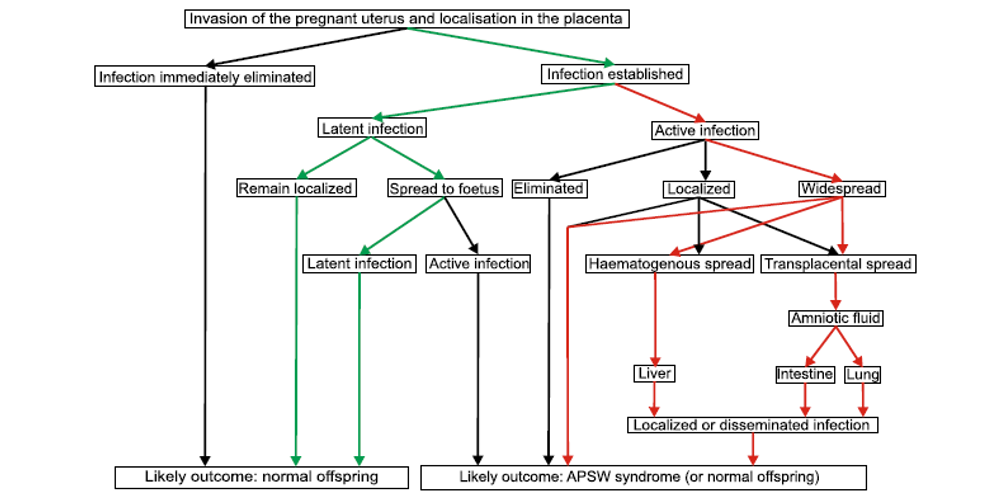
Inhalation of the bacterium, haematogenous spread and then infection of organs. Severity of disease depends on the immunocompetence of the infected animal or human and other factors. In farm animals the bacteria tend to localise in the placenta35. Infected animals will shed the bacteria in a number of body fluids and to varying degrees, this shedding will further contaminate the environment.
In small ruminants the trophoblast cells of the allantochorion are the primary target and there is increasing severity of inflammation. The trophoblasts over the cotyledonary villi are not affected and therefore, unlike EAE or Brucellosis, foetal death is seen shortly before or during abortion or may be born alive (although compromised)15. Generally, there are no symptoms preceding abortion in infected small ruminants.
In pregnant animals a suggestion of pathogenesis is shown below.9
Shedding:
Infected animals can shed the bacteria through a number of routes14,36.
The most common and consistent route is in the placenta and fluids at parturition, with levels of up to 109 bacteria per gram of placenta2, the calving or kidding area can rapidly have a high level of bacteria that may persist for some time.
Guatteo et al (2006)36 investigated 31 herds known to have infection. 242 cows, that had either had a positive PCR post calving or calved within 45 days of an aborted animal or were seropositive, had samples of vaginal mucus, faeces and milk taken to assess the shedding routes.
Overall, 45.4% of the cows sampled were detected to be shedding and this proportion was consistent for both aborted cows and the other in contact or seropositive cows.
Table 1: Which route where the cows shedding from:
| Milk | Faeces | Vaginal mucus | |
| % Positive samples | 24.4 | 20.7 | 19 |
Table 2: Did cows shed from one or more routes:
| One route | Two routes | Three routes | |
| % Positive cows | 65 | 14.6 | 6.4 |
The same author37 demonstrated:
Herds with a negative BM PCR had 7.3% shedders in the herd compared to PCR positive BM herds which had 21% shedders, the study also proved a link between increasing PCR titres and the prevalence of shedder animals.
Shedding can be classified as persistent, sporadic, intermittent or absent.
It also been shown that cows that are seronegative and appear to be healthy can also be shedding.28
Table 3: The percentage of sheders and level of shedding (measured in bacteria per ml of vaginal mucus):
| Overall | Low (0-100) | Medium (101-10,000) | High (>10,000) | |
| Seronegative | 12.7% | 55.4% | 26.1% | 18.5% |
| Seropositive | 26% | 40.2% | 22.7% | 37.1% |
These variations contribute to making a diagnosis challenging (see diagnostic section later).
Clinical signs
- Goats – abortion is the main sign and can have high morbidity. Infection will increase the risk of retained foetal membranes (RFM).
- Cattle – more variable fertility impacts
- Infection can result in abortion, still birth and weak calves
- Increased risk of RFM, metritis and endometritis
- Ordronneau 201210 demonstrated an increased risk of abortion by a factor of 2.5 in seropositive animals and an increased risk of RFM by a factor of 1.5 in seropositive animals when compared to seronegative animals.
- Potential for increased time from calving to conception, decreased CR, increased pregnancy loss
Zoonotic potential
Coxiella burnetii can cause significant disease in humans. Most cases appear to be linked to small ruminant reservoirs. This may be due to burden of infection and contact or could be genetic variation within the bacteria with the small ruminant strains appearing more pathogenic.18
Q Fever Diagnosis
Q Fever is not always included in routine laboratory testing.
In small ruminants it should be considered a differential in abortion cases, especially when you consider that it, like Chlamydia abortus (cause of EAE), will stain with red staining elementary bodies with a Ziehl Neelsen stain. PCR testing the foetal stomach contents (FSC), placenta or vaginal mucosa is a good way to establish if Coxiella burnetii may be involved.
In cattle diagnosis can be more difficult to establish.
- Bulk milk PCR will detect if there is an infected animal shedding into the herd and so demonstrate circulation of the bacteria. It is important to note that shedding can be intermittent but of all the fluids tested milk is the most consistent and persistent so most likely to get an accurate result.
- As with small ruminants, in the case of abortions PCR testing of the FSC or vaginal mucosa will be of use.
- In herds where infection is suspected to be impacting on reproductive performance, a bulk milk PCR may be of use but also ELISA tests on serum can be very useful. Blood sample 6 cows that have had reproductive issues and assess ELISA level. It may also be of benefit to blood sample 6 bulling heifers as these tend to be seronegative and then seroconvert when joining the herd and so this may impact on herd performance.
UK support for testing
Offer the Q Test for BM PCR testing and PCR testing of cervical swabs taken within a week of abortion.
The Q Test uses FTA cards which can then easily be sent to the French Lab for analysis whilst minimising the risk of aerosols (unlike raw BM) which could infect people handling the samples. These can be sent to you and contain everything you need to run this test. Q Test packs can be requested from your Ceva Territory Manager.
The Q Test has recently been validated with the data published in the Journal of Dairy Science38. The DNA remained stable on the FTA card for 28 days and the FTA cards had a higher detection rate at 91.4% compared to raw milk at 77.6%.
Subsidised serology (up to 12 samples per farm) can be submitted to Biobest laboratories. Speak to your Ceva Veterinary Advisor for advice on diagnostics for Q fever.
Prevention and management
Once a diagnosis is made excellent hygiene and biosecurity are important. Treatment with antibiotics is neither considered responsible use of antimicrobials nor is there significant data to support its efficacy.
Goats26
2 injections of 20mg/kg oxytetracycline at 104 qn5 and 120 days of gestation did not reduce the number of abortions or prevent shedding.
Sheep27
A double oxytetracycline treatment at days 100 and 120 of gestation neither prevented the shedding of bacteria nor limited the duration of bacterial excretion. This protocol did not reduce bacterial load excretion nor show any beneficial residual effect in the following lambing season in terms of reducing or eliminating bacterial excretion in previously treated ewes.
Cattle28
An injection of tetracycline at drying off did reduce shedding at calving but repeating the injection had no further benefit and the antibiotic did not reduce the bacterial load. There was large variation in the response to treatment across the herds studied.
Vaccination
COXEVAC® is a phase one inactivated unique vaccine against Q Fever.
Goats29
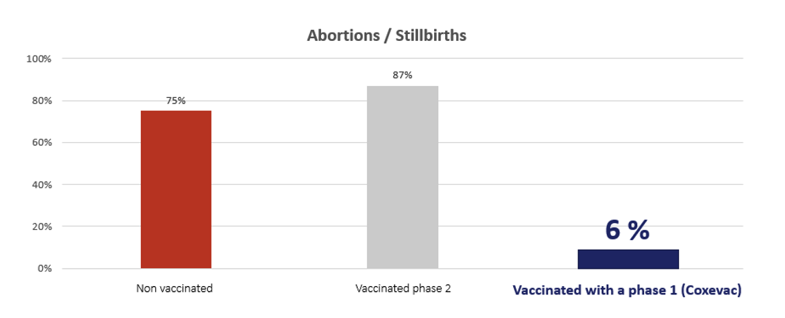
Phase one vaccine had a significant impact in reducing the number of abortions.
The phase one vaccine also had a strong effect in reducing for how long bacterial excretion occurred after kidding.
| Vaginal shedding | Fecal shedding | Milk shedding | |
| Non vaccinate animals | 22 days | 27 day | 17 days |
| Vaccinated with a phase II | 16 days | 28 days | 14days |
| Vaccinated with a phase I | 1.5 days | 10 days | 0 days |
Cattle
Ordronneau10 studied 74 (positive) herds and over 5000 cows and demonstrated that vaccinated cows were 30% less likely to abort when compared to unvaccinated herds.
Guatteo30 (2008) demonstrated that susceptible animals, negative PCR on milk, faeces and vaginal mucus as well as seronegative on 2 blood samples 2 weeks apart, vaccinated pre service were 5 times less likely to become shedders.
A vaccination program put in place for all animals over 3 months of age over a 3 year period reduced the overall number of shedder cows as well as reduced the environmental levels of the bacteria31(Pinero 2014) so reducing the risk of animals being exposed to the pathogen.
Whole herd vaccination reduces the bacterial load across the herd32.
Vaccinated heifers had a reduction by 50% in their risk of having a late return to oestrus10.
Lopez-Helguera (2013)39 demonstrated that in an infected herd there was an increased conception rate of 41.9% in vaccinated animals compared to control animals at 30.1%. The same work demonstrated a decrease in calving to conception time from 106 days in control animals to 92 in vaccinated animals.
Potential cost benefits
Reduce abortion estimated to cost £63040
Reduce open days costed at £4 per day41
Veterinarians
This area has more details on infection with Coxiella burnetii / Q Fever which is an important zoonotic disease with a wide geographical distribution.
Positive Coxiella burnetti PCR results should be reported as notifiable zoonotic disease.
A person who has reasonable grounds to suspect that an animal or animal product is affected, or may be affected with a disease, whether by reason of an examination, test, including a laboratory test or otherwise, shall, without any delay, notify the fact or suspicion to an officer of the Minister at the local office of the Department or in cases where an official control programme is in place, to the persons or authority responsible for that control programme.
More information can be found here.
Coxiella burnetii is an obligate intracellular gram-negative bacterium. It can have 2 different forms33
- The large cell variant (LCV) which is >0.5Um and is metabolically active with exponential reproduction
- The small cell variant (SCV) which is a small rod (0.2-0.5um) that is a stationary non replication form responsible for its impressive environmental persistence.
The bacteria are resistant to wide variations in pH, temperature and desiccation as well as being resistant to many commonly used disinfectants33.
Infection is most commonly acquired through inhalation of dust or aerosols containing the pathogen34.
Pathogenesis:
Inhalation of the bacterium, haematogenous spread and then infection of organs. Severity of disease depends on the immunocompetence of the infected animal or human and other factors. In farm animals the bacteria tend to localise in the placenta35. Infected animals will shed the bacteria in a number of body fluids and to varying degrees, this shedding will further contaminate the environment.
In small ruminants the trophoblast cells of the allantochorion are the primary target and there is increasing severity of inflammation. The trophoblasts over the cotyledonary villi are not affected and therefore, unlike EAE or Brucellosis, foetal death is seen shortly before or during abortion or may be born alive (although compromised)15. Generally, there are no symptoms preceding abortion in infected small ruminants.
In pregnant animals a suggestion of pathogenesis is shown below.9
Shedding:
Infected animals can shed the bacteria through a number of routes14,36.
The most common and consistent route is in the placenta and fluids at parturition, with levels of up to 109 bacteria per gram of placenta2, the calving or kidding area can rapidly have a high level of bacteria that may persist for some time.
Guatteo et al (2006)36 investigated 31 herds known to have infection. 242 cows, that had either had a positive PCR post calving or calved within 45 days of an aborted animal or were seropositive, had samples of vaginal mucus, faeces and milk taken to assess the shedding routes.
Overall, 45.4% of the cows sampled were detected to be shedding and this proportion was consistent for both aborted cows and the other in contact or seropositive cows.
Table 1: Which route where the cows shedding from:
| Milk | Faeces | Vaginal mucus | |
| % Positive samples | 24.4 | 20.7 | 19 |
Table 2: Did cows shed from one or more routes:
| One route | Two routes | Three routes | |
| % Positive cows | 65 | 14.6 | 6.4 |
The same author37 demonstrated:
Herds with a negative BM PCR had 7.3% shedders in the herd compared to PCR positive BM herds which had 21% shedders, the study also proved a link between increasing PCR titres and the prevalence of shedder animals.
Shedding can be classified as persistent, sporadic, intermittent or absent.
It also been shown that cows that are seronegative and appear to be healthy can also be shedding.28
Table 3: The percentage of sheders and level of shedding (measured in bacteria per ml of vaginal mucus):
| Overall | Low (0-100) | Medium (101-10,000) | High (>10,000) | |
| Seronegative | 12.7% | 55.4% | 26.1% | 18.5% |
| Seropositive | 26% | 40.2% | 22.7% | 37.1% |
These variations contribute to making a diagnosis challenging (see diagnostic section later).
Clinical signs
- Goats – abortion is the main sign and can have high morbidity. Infection will increase the risk of retained foetal membranes (RFM).
- Cattle – more variable fertility impacts
- Infection can result in abortion, still birth and weak calves
- Increased risk of RFM, metritis and endometritis
- Ordronneau 201210 demonstrated an increased risk of abortion by a factor of 2.5 in seropositive animals and an increased risk of RFM by a factor of 1.5 in seropositive animals when compared to seronegative animals.
- Potential for increased time from calving to conception, decreased CR, increased pregnancy loss
Zoonotic potential
Coxiella burnetii can cause significant disease in humans. Most cases appear to be linked to small ruminant reservoirs. This may be due to burden of infection and contact or could be genetic variation within the bacteria with the small ruminant strains appearing more pathogenic.18
Q Fever Diagnosis
Q Fever is not always included in routine laboratory testing.
In small ruminants it should be considered a differential in abortion cases, especially when you consider that it, like Chlamydia abortus (cause of EAE), will stain with red staining elementary bodies with a Ziehl Neelsen stain. PCR testing the foetal stomach contents (FSC), placenta or vaginal mucosa is a good way to establish if Coxiella burnetii may be involved.
In cattle diagnosis can be more difficult to establish.
- Bulk milk PCR will detect if there is an infected animal shedding into the herd and so demonstrate circulation of the bacteria. It is important to note that shedding can be intermittent but of all the fluids tested milk is the most consistent and persistent so most likely to get an accurate result.
- As with small ruminants, in the case of abortions PCR testing of the FSC or vaginal mucosa will be of use.
- In herds where infection is suspected to be impacting on reproductive performance, a bulk milk PCR may be of use but also ELISA tests on serum can be very useful. Blood sample 6 cows that have had reproductive issues and assess ELISA level. It may also be of benefit to blood sample 6 bulling heifers as these tend to be seronegative and then seroconvert when joining the herd and so this may impact on herd performance.
Ireland support for testing
Offer the Q Test for BM PCR testing and PCR testing of cervical swabs taken within a week of abortion.
The Q Test uses FTA cards which can then easily be sent to the French Lab for analysis whilst minimising the risk of aerosols (unlike raw BM) which could infect people handling the samples. These can be sent to you and contain everything you need to run this test. Q Test packs can be requested from your Ceva Territory Manager.
The Q Test has recently been validated with the data published in the Journal of Dairy Science38. The DNA remained stable on the FTA card for 28 days and the FTA cards had a higher detection rate at 91.4% compared to raw milk at 77.6%.
Subsidised serology (up to 12 samples per farm) can be submitted to a laboratory in France. Speak to your Interchem Veterinary Advisor for advice on diagnostics for Q fever.
Prevention and management
Once a diagnosis is made excellent hygiene and biosecurity are important. Treatment with antibiotics is neither considered responsible use of antimicrobials nor is there significant data to support its efficacy.
Goats26
2 injections of 20mg/kg oxytetracycline at 104 qn5 and 120 days of gestation did not reduce the number of abortions or prevent shedding.
Sheep27
A double oxytetracycline treatment at days 100 and 120 of gestation neither prevented the shedding of bacteria nor limited the duration of bacterial excretion. This protocol did not reduce bacterial load excretion nor show any beneficial residual effect in the following lambing season in terms of reducing or eliminating bacterial excretion in previously treated ewes.
Cattle28
An injection of tetracycline at drying off did reduce shedding at calving but repeating the injection had no further benefit and the antibiotic did not reduce the bacterial load. There was large variation in the response to treatment across the herds studied.
Vaccination
COXEVAC® is a phase one inactivated unique vaccine against Q Fever.
Goats29
Phase one vaccine had a significant impact in reducing the number of abortions.
The phase one vaccine also had a strong effect in reducing for how long bacterial excretion occurred after kidding.
| Vaginal shedding | Fecal shedding | Milk shedding | |
| Non vaccinate animals | 22 days | 27 day | 17 days |
| Vaccinated with a phase II | 16 days | 28 days | 14days |
| Vaccinated with a phase I | 1.5 days | 10 days | 0 days |
Cattle
Ordronneau10 studied 74 (positive) herds and over 5000 cows and demonstrated that vaccinated cows were 30% less likely to abort when compared to unvaccinated herds.
Guatteo30 (2008) demonstrated that susceptible animals, negative PCR on milk, faeces and vaginal mucus as well as seronegative on 2 blood samples 2 weeks apart, vaccinated pre service were 5 times less likely to become shedders.
A vaccination program put in place for all animals over 3 months of age over a 3 year period reduced the overall number of shedder cows as well as reduced the environmental levels of the bacteria31(Pinero 2014) so reducing the risk of animals being exposed to the pathogen.
Whole herd vaccination reduces the bacterial load across the herd32.
Vaccinated heifers had a reduction by 50% in their risk of having a late return to oestrus10.
Lopez-Helguera (2013)39 demonstrated that in an infected herd there was an increased conception rate of 41.9% in vaccinated animals compared to control animals at 30.1%. The same work demonstrated a decrease in calving to conception time from 106 days in control animals to 92 in vaccinated animals.
Potential cost benefits
Reduce abortion estimated to cost €78240
Reduce open days costed at €4 per day41